What is Alloimmunization & How Can RHD Genotyping Help?
Alloimmunization is an immune response to foreign blood types (antigens). Because the red blood cells of different individuals express many different blood group antigens, exposure to foreign red blood cells, such as in cases of transfusion or pregnancy, can lead to the formation of antibodies. These antibodies bind to red cells expressing the foreign antigen, leading to adverse transfusion reactions. Alloimmunization during pregnancy poses significant risk to the fetus if fetal cells express the corresponding antigen. Blood typing techniques such as RHD genotyping are essential for minimizing the risk of alloimmunization during pregnancy.
Causes of Alloimmunization
Alloimmunization is caused by exposure to foreign antigens, such as receiving a blood transfusion or during pregnancy and delivery. White blood cells recognize the antigen as foreign and mount an immune response against the red blood cells expressing the foreign antigen.
For example, alloimmunization may cause hemolytic disease of the fetus and newborn (HDFN), a condition in which a pregnant woman develops antibodies against antigens expressed on the red blood cells of her fetus/newborn. Although symptoms vary greatly in severity, HDFN can be fatal if untreated. The American College of Obstetricians and Gynecologists (ACOG) estimates about one out of seven fetuses affected by RhD-mediated hemolytic disease are stillborn, with about half of the live births resulting in brain injury or death.
The most clinically significant risk of alloimmunization occurs when the mother’s blood type is RhD-negative and the fetal blood type is RhD-positive. In this case, the mother’s immune system may develop RhD antibodies in response to exposure to fetal red blood cells. Although RhD alloimmunization rarely affects the first pregnancy, a subsequent pregnancy with another RhD-positive fetus is at significant risk of HDFN if the mother does not receive Rh immunglobulin.
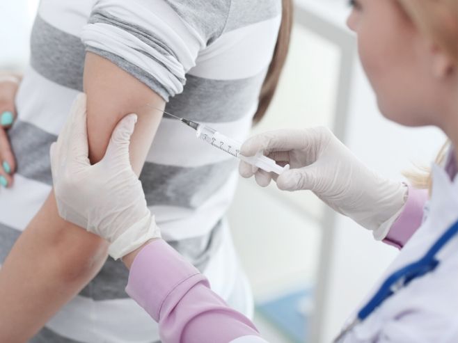
Routine Typing Practices
In routine serological RhD blood typing, the patient’s red blood cell are mixed with anti-D typing reagent. Agglutination of red cells indicates the presence of the RhD antigen (Rh positive), whereas a lack of agglutination means the patient’s cells don’t express the RhD antigen (Rh negative). A more sensitive serologic test employs anti-human globulin reagent to detect the presence of weak reactivity that may be undetectable with direct agglutination testing. This indirect antiglobulin method is referred to as serologic weak D testing. When an individual’s red cells react weakly (<2+) with direct agglutination testing but react moderately or strongly with the addition of antihuman globulin, he/she is said to have a serologic weak D phenotype. Serologic weak D phenotypes are a result of RHD gene variants that affect the expression of the RhD antigen on the red blood cells.
Most laboratories do not perform serologic weak D testing on patients; thus patients with serologic weak D phenotypes are often treated as RhD-negative. For blood donors, on the other hand, weak D testing is performed, and individuals with a serologic weak D phenotype are considered RhD-positive. These differences in testing can easily lead to confusion.
In cases of pregnancy, the administration of Rh immune globulin (RhIG) is the standard treatment to prevent RhD alloimmunization in RhD-negative women. This prophylaxis is 98.4 to 99 percent successful.
RHD Genotyping
The College of American Pathologists (CAP) Transfusion Medicine Resource Committee (TMRC) conducted a survey of more than 3,100 laboratories in the United States in 2014. This survey focused on the policies and procedures for testing and treating patients with serologic weak D phenotypes. The most significant finding of the survey was that there is a lack of standard practice regarding the interpretation of test results in cases of serologic weak D phenotypes, and these differences affect patient care.
As a result, AABB and CAP convened a Work Group, which was charged with the task of developing recommendations for RHD genotyping in patients with a serologic weak D phenotype. The Work Group recommended implementation of RHD genotyping in cases of serologic weak D phenotypes. Furthermore, the Work Group acknowledged that patients with Weak D Types 1, 2 or 3 are not at risk for RhD alloimmunization, and therefore may be managed safely as RhD-positive. This is significant because about 95 percent of Caucasians from central Europe with a serologic weak D phenotype are Weak D Types 1, 2 or 3.
RHD genotyping improves the accuracy of RhD determination. This testing can identify the individuals who are Weak D Types 1, 2 or 3 who can be managed safely as RhD-positive. Thus, RHD genotyping can reduce unnecessary administration of RhIG and transfusions of RhD-negative blood, which is often in short supply.
For more on the benefits of RHD genotyping for prevention of alloimmunization, download “How do I manage Rh typing in obstetric patients?” by Haspel et al. (2015) here.